-
Emergency
-
Apollo Lifeline
![]() Verified By Apollo Doctors December 30, 2022
Verified By Apollo Doctors December 30, 2022
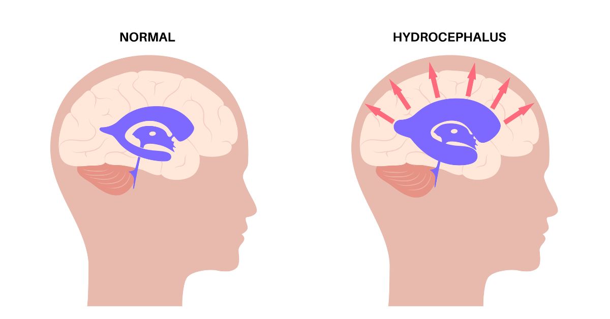
It is known as hydrocephalus when a person suffers from cerebral spinal fluid (CSF) buildup in the brain. The condition increases the pressure in the brain resulting in numerous health conditions. Therefore, doctors recommend inserting the ventriculoperitoneal shunt in the patient’s brain. The shunt is a medical device that drains the excess CSF and relieves the brain from excessive pressure.
This blog explains ventriculoperitoneal shunt, its types, procedure, surgery, and recovery.
A ventriculoperitoneal shunt is a medical device used to treat hydrocephalus – a brain condition where excess CSF accumulates in the brain. Doctors surgically insert this thin hollow tube in the brain, sometimes in the spine, to drain the excess fluid and redirect it into other body parts, where it gets reabsorbed into the blood, thus relieving the pressure and the symptoms of hydrocephalus.
Cerebral spinal fluid cushions and protects the brain from injuries inside the skull. The CSF functions to deliver nutrients to the brain and eliminates waste products. Typically, CSF flows through the ventricles to reach the base of the brain. It cushions the brain and spinal cord before getting reabsorbed into the blood.
However, when the normal flow is interrupted, the CSF accumulates in the brain resulting in harmful pressure on the brain and, eventually, brain damage. The doctor surgically inserts the shunt to relieve the pressure and restore normal flow and absorption of CSF. When the procedure is conducted, the patient is under general anaesthesia. Sometimes, doctors place the shunt in the lumbar region of the spinal cord to restore normal CSF flow and relieve the pressure on the brain. The procedure may provide longer-term benefits.
Two shunts, thin medical, hollow tubes known as catheters, are inserted in a patient’s brain to drain the CSF. The doctor drills a small hole into the patient’s brain and inserts one catheter, which drains the fluid. It is called the inflow catheter. The other catheter placed under the skin ensures the excess fluid gets drained to some other body part. The catheter is known as the outflow catheter. A valve also present, called the pump, controls the shunts in draining the fluid as required.
There are two types of ventriculoperitoneal shunts. They are as follows:
Anyone, irrespective of their age, can develop hydrocephalus. Therefore, patients need a ventriculoperitoneal shunt. The experts of the Mayo Clinic believe that babies and older adults are more likely to develop hydrocephalus. The National Institute for Neurological Disorders and Stroke evaluates that one to two in 1,000 babies are born with hydrocephalus. Also, the Hydrocephalus Association believe that over one million people in the United States have developed the condition. The reasons for fluid buildup are many, and they are as follows:
However, blockages are the primary and common cause of excessive fluid accumulation in the brain. Other causes may include head trauma, stroke, infection, cysts, tumours, or brain inflammation. The person experiencing hydrocephalus may experience the following symptoms:
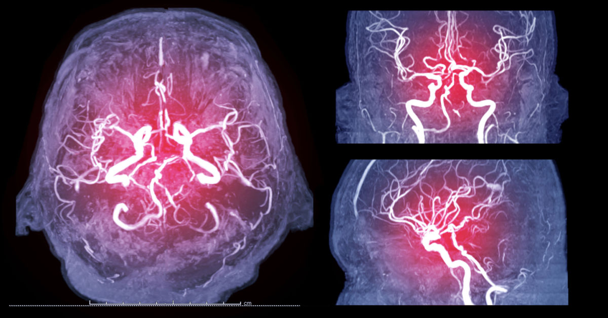
Doctors use various imaging tests such as ultrasound, CT scans, and MRI scans that help the healthcare provider view the brain’s cavities and tissues and help diagnose the condition. These tests show the regions where there is excessive fluid in the brain.
The shunts may help improve the symptoms of hydrocephalus within days of shunt insertion, or sometimes, it may take weeks or months to see noticeable changes.
Nearly 10 per cent of patients with shunts have shown noticeable improvement with the placement of the shunt but with a low long-term response. The cause for this is unknown to the doctors.
It is essential to speak or consult the surgeon before the procedure to inform the surgical team about the medications, supplements, vitamins, or herbs the patient consumes. The surgical team speaks to the patient about preoperative food and drink limitations. Doctorswill need older children and adults to fast at least eight hours before the surgery. However, parents of babies and toddlers must stop feeding their children formula or solid food six hours before the surgery. Only a healthcare provider or a surgical team member should instruct the patient or the patient’s caretaker. The patient should disclose the following information before the procedure:
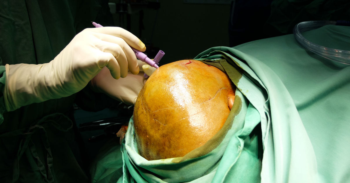
The doctor administers general anaesthesia before performing the ventriculoperitoneal shunts. Once the patient is anaesthetised, the surgical nurse shaves the area behind the ear – this is in preparation for the procedure.
Catheters are flexible, thin tubes used to drain excess fluid. The surgeon will make a tiny cut behind the ear and also drill a small hole in the skull. Then, the surgeon will thread one catheter into your brain through this opening and the other catheter goes behind the ear and is subcutaneous (meaning it resides under your skin). This tube travels down to the chest and abdomen, allowing excess CSF to drain into your abdominal cavity, where the body absorbs it.
The surgeon may attach a tiny pump to both the catheters and place it under the skin behind the ear. The pump, also called a valve, will activate automatically to remove fluid when the pressure in the skull raises. It may also be possible to program the pump to activate when the fluid increases to certain volume.
After all the parts of the shunts are correctly connected, the shunts immediately begin to drain the excess fluid from the brain. Doctors perform the entire procedure in about 90 minutes.
After the completion of the procedure, the patient must lay flat for 24 hours. The patient may experience mild headaches, for which the physician provides pain medications. The patient’s hospital duration is dependent on the reason for the shunt. The hospital staff and the surgical team closely monitor the patient’s blood pressure and heart rate, feeds the patient fluids intravenously, and administer antibiotics and other necessary medications. The patient may be required to stay in the hospital for at least seven days before discharge.
Before going home, the doctor provides clear instructions to the patient on postoperative shunt care at home, taking prescription medications, diet, and follow-up checks. The doctor also checks if the shunts are efficiently functioning before the patient leaves the hospital.
Consult the doctor immediately or visit the nearest hospital if a patient experiences the following signs:
Soon after the surgery, the patient is advised to start with liquids before gradually starting solid foods. During the follow-up visit, the patient’s stitches are removed, incisions are cleaned, and checked for signs of infection. The following are the signs of infection:
The patient is also instructed on when to start showering again, as they cannot begin to shower soon after the surgery due to the incision getting wet and affecting the healing process. Resting well aids in better recovery. Daily activities and work should be resumed only after a doctor’s consultation.
In some cases, if shunts malfunction, they may require replacement. A malfunctioning shunt may either over or under-drain the CSF. If it underdrains, the symptoms of hydrocephalus may reoccur. However, it damages the brain and causes brain haemorrhage if it over drains.
Sometimes, the shunt can be infected. The following are signs of infection:
Once the shunt insertion is done, the patient should avoid being in the presence of a strong magnetic field as it may affect the functioning of the shunt. The patient must inform the radiologist about the shunt during future MRI scans. Earphones may also affect the efficient functioning of the shunt. Therefore, read the shunt’s manufacturing guidelines before using an earphone.
A blockage is another common complication that can be easily fixed and may result in serious harm. Most doctors recommend changing shunts every six years.
Shunting successfully reduces pressure in the brain in maximum number of people. VP shunts may need replacement after many years, particularly in small children. The average lifespan of a shunt in an infant is two years. Children and adults over the age of 2 may not require a shunt replacement for eight or more years. Shunt systems need frequent monitoring and follow-up.
At Apollo, we believe that easily accessible, reliable health information can make managing health conditions an empowering experience. AskApollo Online Health Library team consists of medical experts who create curated peer-reviewed medical content that is regularly updated and is easy-to-understand.


July 7, 2023