Home Treating A Trauma Patient in The ER

      Treating A Trauma Patient in The ER

      Cardiology Image 1 Verified By February 25, 2020

      4750
      Treating A Trauma Patient in The ER

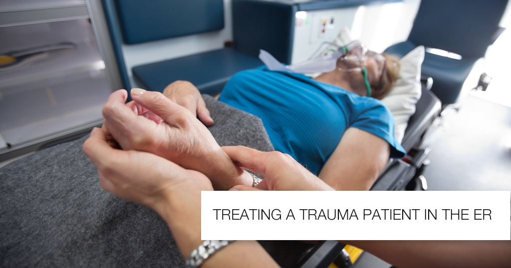
      Treating a trauma patient in the ER starts with a brief but complete handoff from the EMS Team regarding the Pre-Hospital Care received by the patient.

      Pre-Hospital Trauma Care

      Prehospital care of trauma patients varies based on the patient and magnitude of trauma but always focuses on stabilization of the patient and prompt transport to a hospital. The ABCDE approach is the most commonly used by EMS Teams on the scene, albeit in its abbreviated form for the benefit of time.

      Life-Saving Interventions that may be performed by the EMS Team before transporting to a hospital may include, but are not limited to:

      • Placement of a cervical collar (if C-Spine trauma is suspected based on Primary Survey or mechanism of injury)
      • Oxygen delivery via nasal cannula or Intubation (if respiratory distress or unprotected airway)
      • Administration of IV fluid (if hemorrhage or hypotension)
      • Administration of Analgesia
      • Placement pressure bandages for control of bleeding

      Primary Survey (Advanced Trauma Life Support)

      Primary Survey consists of 5 steps that are as follows:

      AIRWAY (With C-SPINE Stabilization)

      • If appropriately answering questions, patient has a patent airway.
      • Observe patient for signs of respiratory distress.
      • Inspect mouth and larynx for injury or obstruction.
      • Assume C-Spine injury in trauma patients until proven otherwise.
      • If patient is unconscious or in respiratory distress, plan to Intubate early.
      • Plan to intubate or ventilate with the anterior portion of the cervical collar removed, or with the neck manually stabilized.
      • Patients with burn injuries and evidence of respiratory involvement should be intubated early.
      • If difficult, perform a Cricothyrotomy.

      BREATHING

      • Assess oxygenation status with Pulse Oximetry.
      • Inspect, percuss and auscultate the chest wall for injuries.
      • Do not delay treatment of Tension Pneumothorax or hemothorax in favour of imaging.

      CIRCULATION (With Hemorrhage Control)

      • Assess the circulatory status quickly by palpating the central and peripheral pulses.
      • Blood pressure should be measured immediately but should not hinder the progress of the Primary Survey if so, it can be delayed.
      • IV access should be promptly secured with two large-bore IV lines(at least 16 gauge) for IV fluids, blood grouping, typing and crossmatch, and resuscitation (if needed).
      • If intravenous line placement is not possible or difficult, intraosseous line should be used instead.
      • Control on-going hemorrhage with manual pressure or tourniquets.
      • If hypotensive, administer a bolus of IV crystalloids.
      • If the history of hemorrhage or on-going hemorrhage, plan and execute immediate Massive Transfusion Protocol.
      • If significant hemorrhage and persistent hemodynamic instability ensue, transfuse Plasma, Platelets and Packed RBCs at 1:1:1 ratio.
      • Focussed Assessment with Sonography for Trauma or FAST examshould be performed, especially for hemodynamically unstable patients.
      • Check for External Hemorrhage.
      • Always rule out Internal Hemorrhage: Thoracic, Pelvic and Abdominal cavities, Thighs

      DISABILITY (With Neurological Evaluation)

      • Assess the patient’s Glasgow Coma Scale score. GCS < 8 is an indication for intubation.
      • Assess both pupils.
      • If patient is interactive, assess motor function and light touch sensation.

      EXPOSURE (With Environmental Control)

      • Undress patient completely.
      • Examine body for signs of occult injury, including patient’s back.
      • If patient is hypothermic, cover with warm blankets and infuse warm IV fluids.
      • Palpate for vertebral tenderness and rectal tone.

      Diagnostic Tests

      The choice of diagnostic imaging modality depends on clinical judgment and mechanism of injury. The decision to perform any diagnostic test must be taken after accounting for the patient’s hemodynamic stability and must be weighed against the need for urgent transfer or operative intervention.

      X-Rays

      • Typically acquired after the Primary Survey.
      • Screening x-rays of the C-Spine, Chest, and Pelvis are usually performed but may be skipped if a CT-scan will be performed. Patients with penetrating injuries to the abdomen or thorax is an exception in which, a chest x-ray should always be taken even if a CT-scan is performed.

      FAST exam

      • Typically acquired during the Primary Survey (especially for hemodynamically unstable patients)
      • An extended version (E-FAST) may alternatively be performed, which allows for detection of pneumothorax and Hemothorax.

      CT scans

      • Typically performed after the Primary Survey if the patient is hemodynamically stable (risk of decompensation while inside the scanner, which could be catastrophic)

      Laboratory Investigations

      Lab tests include, but are not limited to:

      • CBC
      • Renal And/or Metabolic Profiles
      • Prothrombin Time
      • Urinalysis
      • Urine pregnancy test (on all women of child-bearing age)
      • Blood glucose
      • ABG ( if associated with shock)

      Secondary Survey

      • Performed after the Primary Survey has been completed and the patient is deemed stable.
      • Includes complete history and thorough physical examination.
      • Additional diagnostic tests are tailored to remaining symptoms, mechanism of injury, and patient comorbidities.
      • The main goal is to minimize the risk of missed injuries.

      Tertiary survey

      • Delayed re-examination of the patient (usually 24 hours after admission)
      • Main goal is to detect changes due to previously undetected injuries.

      Cardiology Image 1

      Related Articles

      More Articles

      Most Popular Articles

      More Articles
      © Copyright 2024. Apollo Hospitals Group. All Rights Reserved.
      Book ProHealth Book Appointment
      Request A Call Back X