The last century has seen several medical advancements and as a result, humans are healthier and with longer lifespans than ever before. Diseases like smallpox that have resulted in millions of deaths have been completely eradicated with vaccinations while diseases like tuberculosis are now treatable with antibiotics. On the other hand, a healthy diet and lifestyle habits have helped morbidity and mortality from several diseases like diabetes, heart attacks, and strokes.
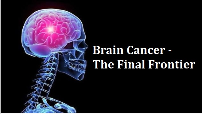
While we live in a very optimistic healthcare environment, there is still risk with some diseases that still have rather grim outlooks, particularly primary brain cancer.
Tumours of the Brain
Brain tumours can be of several types and can affect both children and adults. Tumours of the meninges, which is the covering of the brain, are usually benign and can be treated completely with surgery. In children, tumours can be benign or cancerous, but generally have a better prognosis as compared to cancers that affect adults.
Tumours that begin in the brain cells that support and nourish the nerve cells (glial cells) are known as gliomas and generally have poor outcomes. Graded on a scale of 1 to 4, based on the degree of malignancy, glioblastoma (grade 4) is the most common among these and has the worst outcomes. On average, glioblastomas affect 5 out of 1,00,000 people and have a dismal median survival or about 9 to 14 months, even with the best treatment.
Pathology for Cancer Diagnosis
The push to find a way to cure for this disease has never been greater than it has been in the last decade. Cancer is caused by an unregulated proliferation of cells which manages to bypass all the checks and balances that are in place in a body to stop an uncontrolled multiplication of cells. This situation is generally triggered by a mutation or change in the body’s genetic code, which can affect one gene (in certain blood cancers) or multiple genes (like in colon or brain cancer).
Pathology is till date the foundation of cancer diagnosis and includes recognising malignant cells on a slide that is prepared from a biopsy specimen. If a patient has abnormal results in a brain scan are told to undergo a radical excision or a biopsy that is guided by a CT or MRI scan. A sample of the tumour is examined by a pathologist who gives the final diagnosis. Different grades of the tumour can co-exist in glioma and the outcome is generally determined by the highest grade of the tumour. It is not possible to know or predict the location of the tissue that would represent the highest grade of the tumour via a Ct scan or MRI, it may be possible that the biopsy sample does not represent the tumour. In certain kinds of tumour, in inexperienced hands, the error rate for a biopsy may be as much as 30%. Such errors can be avoided with a liquid biopsy.
Liquid Biopsy
Nano and microvesicles, or exosomes which are released by live cells; circulating tumour DNA which is released by dead cells, and tumour cells are released by brain tumours straight into the bloodstream and the cerebrospinal fluid. Isolating and analysing these substrates in the blood offers more accurate diagnosis and also helps identify cancer-causing pathways. It is a fact that the quantity of exosomes being released increases exponentially with each grade of the tumour, so even a small high-grade lesion, which is a low-grade tumour, may be missed with a conventional biopsy, but will be apparent in the liquid form. The MIT voted liquid biopsies as one of the breakthrough technologies of 2015, and the use of the same for brain tumour diagnosis was demonstrated by Apollo Research and Innovations, Hyderabad, a couple of years ago in a published study.
Genetic Tumour Markers
A limitation of pathology is that it provides a diagnosis, but the architecture of the cells does not tell a lot about the possible mutation pathways that are causing cancer. Today, many of the pathways can be targeted with specific drugs to halt the spread of cancer. Recently, the WHO has entrusted an international group of pathologists for the revision of the pathological grading system, and to introduce a genomic component to the diagnosis. This will help improve the accuracy of the diagnosis and can also guide the possible treatment. In the last 5 years, 5-6 genetic markers have been made mandatory for diagnosing a glioma. With the technology available today, it is possible to simultaneously test 30,000 different gene mutations in a given tumour sample. With the addition of more genomic data points in the future, the prognostication and treatment of glioma patients are surely going to improve.
 The Treatments:
- Radical Surgery
- Radiation Therapy
- Chemotherapy
Radical Surgery
An important procedure that can improve the chances of survival in high-grade tumour cases, radical surgery is only considered to be useful if it does not induce paralysis in the opposite side of the body. ‘Awake’ anaesthesia and intra-operative MRI are two technologies that have made quite a difference to the outcomes of radical tumour surgery.
One of the main problems in removing such lesions is that surgeons are unable to predict the ‘edge’ of the tumour, which even under modern operating microscopes appear like the normal brain. Straying away from the edge can cause paralysis while not removing the edge decreases the chances of survival. Awake anaesthesia allows the surgeon to wake the patient up in the middle of the surgery with the tumour exposed, without them feeling any pain. They are awake enough to be able to talk and move their limbs and can tell the surgeon if they are able to move their limbs or if they feel anything abnormal as the surgeon works on removing the tumour. The surgery can be stopped if there is any hint of trouble, which reduces the mortality rates significantly. Intra-operative MRI is another technique that helps in the identification of the edge of the tumour in real-time. When both awake anaesthesia and intra-operative MRI are used in conjunction, the outcomes of radical surgery are significantly improved.
Radiation Therapy
Radical surgery or biopsy (for patients who have inoperable tumours) is considered to be the first step of treatment and radiation surgery is important for these patients. A high dose of radiation is administered by an external beam which damages the DNA of the tumour and causes them to die at the time of replication. The cells keep getting destroyed even long after the radiation therapy has ended. The problem here is that the radiation not only damages cancer cells but also normal tissues that surround it. However, some newer techniques have emerged, which help reduce the side effects of radiation therapy.
Stereotactic radiosurgery is a technique that focuses hundreds of sub-clinical X-Ray beams at one point within the tumour. This maximises the dose being delivered but minimises the exposure of the tissues surrounding the tumour, thereby protecting the adjoining normal parts of the brain from the bad effects of radiation.
Conventional radiation used electrons, however, in recent times, the use of a proton beam has revolutionised radiation therapy for brain tumours in certain situations. Protons are larger particles, carry more energy, and can be focused more accurately within the tumour, as compared to electrons. In this case, the entire energy of the proton is delivered at the tumour bed which means that there are no effects of radiation beyond the tumour. Such accuracy is not possible with conventional radiotherapy.
Chemotherapy
Chemotherapeutic agents have not been at the frontline of managing brain tumour till now but that is something that may change soon in upcoming times. It is a well-kno0wn fact that glioma subgroups with specific mutations show high chemo-sensitivity. Chemotherapy can give some spectacular results if the glioma being treated harbours particular targetable mutations. As the collective knowledge about the biology of cancer keeps increasing, new targets can be discovered.
There are more than 200 clinical trials that are being held all over the world and are recruiting glioma patients in an effort to identify and block cancer-causing pathways in gliomas. There are newer forms of chemotherapy (immune checkpoint inhibitors) that use the body’s immune system to target the cancer cells. The first of such drugs has been now approved by the FDA. This is a significant move towards better brain tumour management in the future.
Today there are a lot of reasons for one to be more optimistic than any time before that we are closer than ever to curing cancer. There have been several improvements in surgical and radiation techniques taking the 3-4 months’ survival rate from 30 years ago to almost 14 months today. Gliomas constitute 1% of all cancers and 3% of cancer-related deaths, which emphasises the advances in the treatment of other cancers. There have been major strides in understanding the biology of cancer and an important part of the strategy has been teasing apart the subgroups that respond to therapy. Hopefully, it won’t be too long before this frontier too is conquered.
To know more about the “Medical Oncology”, watch the below-mentioned video by Dr. P K Das, from Apollo Hospitals, New Delhi.


 The Treatments: