


- Imaging
- Laboratory
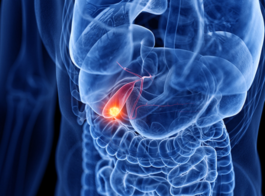
- Ultrasound– is used for detecting the location of tumor and whether the tumor involves the main blood vessels. It can also distinguish whether a mass is cancerous or benign. E.g. Endoscopic or laparoscopic ultrasound.
- Endoscopy- It is a procedure that helps to look inside the throat, esophagus, stomach, and duodenum. An endoscope is a thin, tube-like instrument with a light and a lens for viewing and is inserted into the mouth to examine the interior of the esophagus (food pipe) and stomach. It may also be used to remove tissue samples for testing.
- CT scan/ Contrast CT Scan is a diagnostic imaging procedure that uses x-rays to build cross-sectional images of the gallbladder, liver, bile ducts, and nearby lymph nodes during three phases of blood flow through the liver. It also helps to identify the spread of cancer.
- CT Angiography-Gallbladder is located close to vital structures like hepatic artery and portal vein. Hence tumors of gallbladder can invade these structures. CT abdomen with angiography is done to examine the extent of disease and rule out the involvement of these critical structures before undertaking the surgery.
- Magnetic resonance imaging (MRI) – uses the interaction of radio waves and magnetic field which is processed in a high speed computer system to produce detailed scan pictures of the tissue, organs, bones, ligament and cartilage. It may be useful in detecting tumors and their metastases. This diagnostic technique offers greater soft tissue contrast than a CT scan.
- Magnetic Resonance Cholangiopancreatography (MRCP) – shows how much a tumor has grown within the gallbladder. It helps determine if the tumor can be removed by surgery, and to see if the tumor has spread to the liver or other organs.
- PET CT – is considered to assess spread to regional nodes or distant metastases to other parts of the body. It provides functional and morphological details by utilizing radiation derived from Isotope labeled Glucose molecules to detect cellular glucose uptake, in cancer.
- Ultrasound guided Biopsy of the gallbladder tumor-A small tissue is taken from the gallbladder tumor for the confirmation of diagnosis by microscopic examination (Pathology). The biopsy can be obtained by image (USG/CT) guidance or through laparoscopic surgery. If the surgeon deems the tumor could be removed by surgery, they directly proceed to surgical resection.
