• Emergency
  • Apollo Lifeline

Emergency

Best Doctors for Retinal Detachment in Delhi

Retinal detachment is a severe eye condition where the light-sensitive tissue lining the interior of the eye detaches from its normal position. This often leads to vision loss if not promptly addressed. It can be caused by various factors such as ageing, extreme nearsightedness, previous eye surgery, and eye injuries.

In Delhi, many residents consult retinal detachment doctors due to sudden visual disturbances or loss of sight. Apollo Hospitals’ team of retinal detachment treatment doctors ensure that patients receive timely and effective treatment. The retinal detachment surgeons are known for their expertise and commitment to patient care.

Search Result: 5

Dr Aniel Malhotra

MBBS, MS, DOMS

Registration No

205413

Language

English, français, हिंदी

26 years experience overall

Sarita Vihar , Delhi


TUE, FRI, SAT | MON, WED, THU(12:00 PM-04:00 PM | 10:00 AM-12:00 PM)
Call Now Call Now

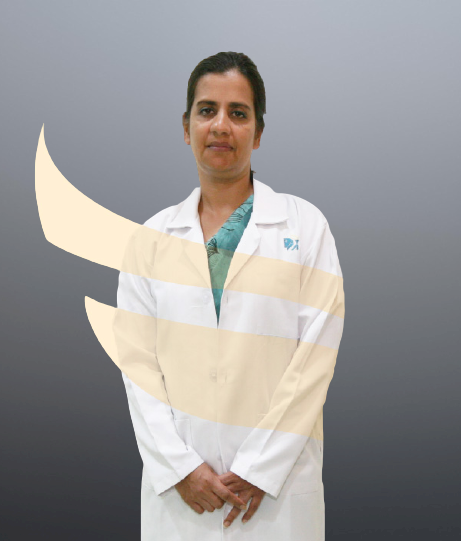
Registration No

678240

Language

English, हिंदी

24 years experience overall

Sarita Vihar , Delhi


THU | SAT | TUE(12:00 PM-04:00 PM | 04:00 PM-06:00 PM | 10:00 AM-12:00 PM)
Call Now Call Now

Dr L R Seth

MBBS, MS, DOMS

Registration No

452

Language

English, हिंदी

30 years experience overall

Sarita Vihar , Delhi


MON, THU, FRI | TUE, WED, SAT | MON, THU, FRI(08:00 AM-10:00 AM | 12:00 PM-04:00 PM | 10:00 AM-12:00 PM)
Call Now Call Now

Registration No

460

Language

English, हिंदी

31 years experience overall

Sarita Vihar , Delhi


MON, FRI | TUE, WED, SAT(12:00 PM-03:00 PM | 10:00 AM-12:00 PM)
Call Now Call Now

Dr Uma Mallaiah

MBBS, DO, FRCS

Registration No

457

Language

English, हिंदी

20 years experience overall

Sarita Vihar , Delhi


TUE, FRI | WED, THU | MON, SAT(10:00 AM-02:00 PM | 01:00 PM-03:00 PM | 02:00 PM-04:00 PM)
Call Now Call Now

Frequently Asked Questions for s in

It is recommended to sleep facing down after retinal detachment surgery. Your doctor may provide further guidance based on your specific situation.

No, flying does not transform retinal holes into detachment. However, changes in cabin pressure during flights can potentially worsen existing retinal conditions. If you have a history of retinal holes or detachment, it is advisable to consult with your ophthalmologist before flying to ensure it is safe for you.

The haloing visionary effect around lights after retinal detachment surgery may be due to temporary changes in the cornea (the clear front part of the eye). These changes can affect how light enters the eye and may cause halos or glare around bright lights. This symptom usually improves over time as the eye heals.

Yes, there is a risk of retinal tears or detachment recurring even after successful treatment. Regular follow-up appointments with your ophthalmologist are essential to keep track of your eye health and detect any signs of recurrence early on.

In general, multiple attempts can be made to reattach a detached retina. However, the success rate may decrease with each subsequent surgery. The number of attempts depends on various factors, including the condition of the retina, the underlying cause of detachment, and the overall health of the eye.

After retinal detachment surgery, common symptoms of recovery may include mild discomfort, redness, and temporary vision changes. These symptoms should gradually improve over time. However, if you experience severe pain, worsening vision, or any unexpected symptoms, it is essential to contact your doctor immediately.

In some cases, a detached retina may be treated with non-surgical methods such as laser therapy or cryotherapy. However, these treatments are generally considered temporary measures and may not be suitable for all cases. In most cases, surgical intervention is necessary to reattach the retina and restore vision.

After retinal detachment surgery, you may need to refrain from activities that put excessive strain on your eyes, such as heavy lifting or strenuous exercise. It is key to follow your doctor’s instructions to ensure healing.

The success rate varies depending on several factors, including the type and severity of detachment, the surgical technique used, and the individual patient’s overall eye health.

You should see a doctor immediately if you experience sudden changes in vision, such as the appearance of floaters (tiny specks or flashes of light), darkening or blurring of vision, or a curtain-like shadow across your visual field. These could be signs of retinal detachment and require urgent medical attention.

Various factors can be responsible for retinal detachment. These include ageing, eye injuries, certain eye conditions like diabetic retinopathy, high myopia (nearsightedness), and previous eye surgeries. In some cases, retinal detachment may occur without any identifiable cause.

Retinal detachment is diagnosed through a comprehensive eye examination. Your ophthalmologist will use various instruments to examine your eyes and may perform additional tests such as ultrasound or optical coherence tomography (OCT). These tests help determine the extent and location of the detachment.

Ophthalmologists, specifically those who specialize in retina diseases (retina specialists), are the specialists who treat retinal detachment. They have advanced specializations in conditions affecting the retina.

While it is not always possible to prevent retinal detachment, there are some steps you can take to reduce the risk. Regular eye examinations, especially if you have a family history or are at increased risk, can help detect any early signs. Prompt treatment of eye injuries or conditions that can lead to detachment can also minimize the risk.

The treatment options for retinal detachment include laser therapy, cryotherapy (freezing treatment), and surgical procedures such as pneumatic retinopexy, scleral buckle surgery, and vitrectomy.

Types/Stages of Retinal Detachment

Retinal Detachment has three main types which are:

  • Rhegmatogenous: This is the most common type and occurs when a small tear or hole in the retina allows fluid to seep behind it and prompt its detachment from the back of the eye.

  • Tractional: Scar tissue on the surface of the retina pulls it away from the back of the eye. This type is often associated with diabetic retinopathy in individuals with poorly controlled diabetes.

  • Exudative: This type happens when fluid builds up under the retina, causing its detachment. However, there are no tears or breaks in the retina.

Symptoms of Retinal Detachment

The symptoms indicating Retinal Detachment include:

  • A sudden increase in floaters and flashes of light

  • Blurred or reduced vision

  • Gradually reduced peripheral (side) vision

  • The appearance of a curtain-like shadow over part of the visual field

Tests to Diagnose Retinal Detachment

At Apollo Hospitals, Delhi, several tests are used by doctors to diagnose retinal detachment. These include:

  • Ophthalmoscopy: The doctor uses a bright light and special lenses to scrutinise the retina for tears, holes, or detachment.

  • Ultrasound imaging: If bleeding obscures direct examination of the retina, ultrasound imaging is utilised to create pictures of the eye’s structure.

  • Amsler grid: Patients use this grid of horizontal and vertical lines to determine any distortion or wavy appearance, indicative of retinal detachment.

Treatment or Surgery Options for Retinal Detachment at Apollo Hospitals in Delhi:

The treatment approaches for retinal detachment at Apollo Hospitals in Delhi include:

  • Laser surgery (photocoagulation): The surgeon uses a laser to create burns around a retinal tear, facilitating the sealing of the retina back into place.

  • Freezing treatment (cryopexy): The surgeon applies a freezing probe to the outer surface of the eye over the retinal tear, creating a scar that secures the retina.

  • Pneumatic retinopexy: In this procedure, the surgeon injects a gas bubble into the eye that pushes the retina back into its normal position.

  • Scleral buckling surgery: The surgeon makes a small indent in the white part of the eye to relieve traction on the retina, allowing it to reattach.

  • Vitrectomy: The surgeon extracts the vitreous gel from the eye and may also drain fluid, inject gas or silicone oil, and perform other procedures to reattach the retina.

© Copyright 2024. Apollo Hospitals Group. All Rights Reserved.

+91 8069991061 Book Appointment

Request A Call Back

X